All About Prostate Cancer
What are the symptoms?

There are normally no symptoms associated with the early-stage prostate cancer. However, as the disease progresses and the tumour gets bigger it may compress and constrict the urethra (the tube which runs through the gland), obstructing the flow of urine during urination.
In this situation the patient may notice a weak, interrupted stream of urine that requires straining to urinate and to empty the bladder completely. On completion he may still feel that the bladder is not empty.
However, these symptoms are not specific to prostate cancer and are most commonly found in benign non-cancerous enlargements of the gland. (This is known as benign prostatic hyperplasia or BPH.)
Blood in the urine or semen may also be a sign of prostate cancer, although again, this is a common finding and not often not related to malignancy.
If a tumour has spread to the bones, then it may cause pain. The spine is the most common site for this to occur giving rise to unexplained back pain.
How is prostate cancer diagnosed?
The Specialist will initially ask the patient questions to check their general medical health, their family history and see if they are experiencing any symptoms associated with prostate cancer (although, as has been mentioned, such symptoms are not specific to prostate cancer).
Medical examination
During a visit to the doctor, a male patient over the age of 50 may be asked questions about his general medical health and whether he has any specific symptoms. The answers that are given may prompt the doctor to carry out further tests to see if there is a possibility that the patient may have prostate cancer.

Physical examination
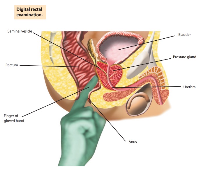
Having made a general examination, the GP or specialist, will then request to perform a Digital Rectal Examination (DRE) to feel the gland. A gloved, lubricated finger is inserted into the back passage (rectum) to check the size and shape of the prostate gland.
Blood test
The prostate can be evaluated by testing for the level of a particular protein in the blood called PSA (prostate-specific antigen). Prostate enlargement tends to cause an increase in the level of PSA, with malignant tumours (cancers) producing a greater increase than benign enlargements. However, other conditions can also cause PSA to rise, such as a urinary infection. Therefore, although a slight elevation in the PSA may indicate prostate cancer, it is by no means definite. Most Patients will therefore be referred in due to an elevated PSA and/or an abnormal DRE.

MRI scan
MRI (magnetic resonance imaging) has changed the way we are diagnosing prostate cancers. If your GP has a suspicion of prostate cancer due to a raised PSA, an MRI scan will be arranged at your local hospital. MRI scans have greatly improved our ability to detect prostate cancer and if your MRI is normal you may not need further tests and can be reassured. MRIs are scored using a system called PIRADS (Prostate Imaging, Reporting and Data System) which ranges from 1 to 5 based on the chance of prostate cancer being present, and usually only prostates scoring 3 or more require a biopsy.
Ultrasound examination and transperineal prostate biopsy
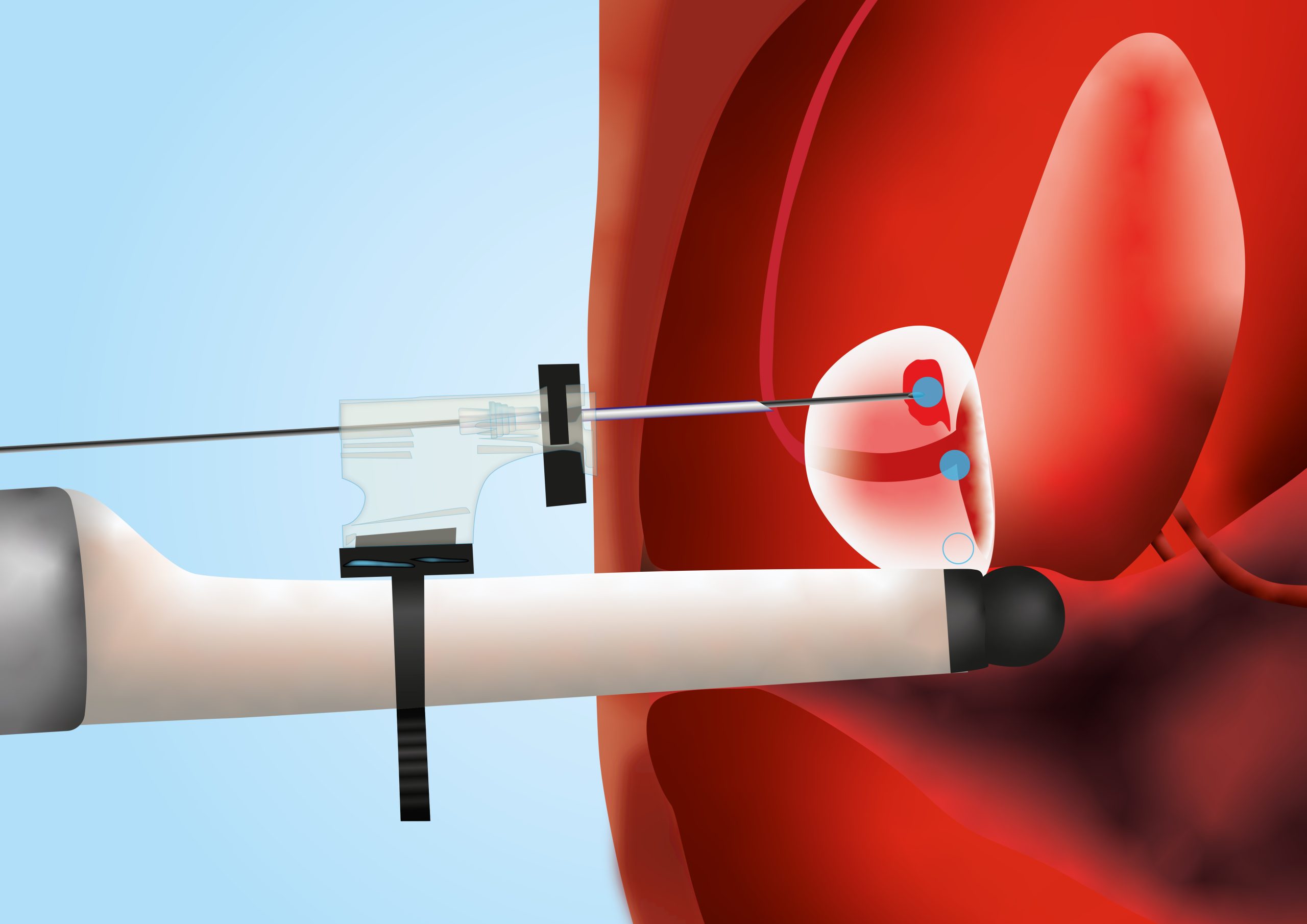
Prostate tissue samples are taken by fine needles passed through the skin that lies between the anus and testicles (the perineum). Local anaesthetic is injected into the perineum and around the prostate prior to biopsies being taken. The procedure is no more painful than giving blood, and antibiotics will be given to minimise the risk of developing infection. Typically, about 20-30 biopsies are normally taken, which are then analysed in the laboratory, and a diagnosis obtained. After the procedure, it is quite common for the patient to see some blood in their urine and semen, but this usually settles over a week or two.
If your MRI suggests you need a biopsy of your prostate, this is may be performed under a local or general anaesthetic as a day case procedure. An ultrasound probe similar in size to a finger, is inserted into the rectum, and images of the prostate appear on a screen. These images in conjunction with the MRI images will help your specialist to target the suspicious areas in the prostate.
Bone scan
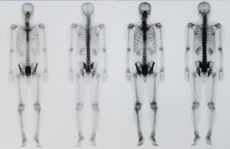
Once a diagnosis of prostate cancer has been made, if spread is suspected (usually by the level of PSA and/or grade of cancer), one of the imaging tests that can be use is a bone scan, used to see if the tumour has spread to the bones. For this painless test, a tiny, harmless quantity of a radioactive agent is injected into a vein. This makes its way to any cancer deposits within the skeleton and sticks to them. After a few hours, the patient is scanned by a special camera, similar to an x-ray machine, which detects these deposits, if present.
PSMA-PET/CT scan
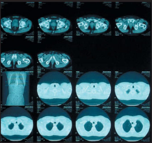
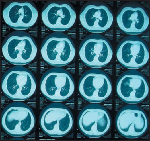
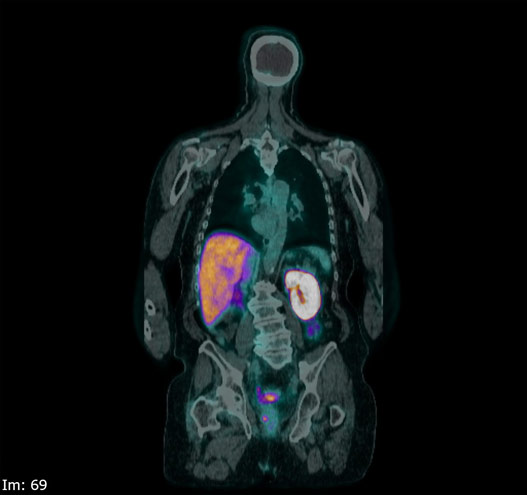
For patients with higher risk prostate cancer, a PSMA PET/CT scan may be performed. This can detect spread of cancer into the lymph nodes in the body or bony skeleton. It is a specialist scan again involving injection of a tiny, radioactive agent PSMA (Prostate Specific Membrane Antigen) into the blood that sticks to cancer deposits wherever present in the body. These are then highlighted on the images.
Overall cancer assessment
Once all diagnostic information is available, patients are usually reviewed in a multidisciplinary team (MDT) meeting, (with Urologists, Oncologists, Specialist nurses, Pathologist and Radiologist) to verify the findings and to consider the suitable treatment options. Often, patients are divided into risk-group categories to help determine which treatment they are best suited to. There are different ways to categorise patients based on the risk of the cancer spreading and causing harm. These include the Cambridge Prognostic Groups (CPG), ISUP and NICE risk stratification.
Grades of prostate cancer
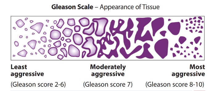
The pathological grade of a cancer is the term used to describe how aggressive the disease is and whether it will progress (grow) quickly (months) or slowly (years). The grading assessment is made by a pathologist in the laboratory, looking at cells from the biopsy sample under the microscope. If cancer cells are found in the biopsy samples, the pathologist will ‘grade’ them. The grading system used for prostate cancer is known as the Gleason scoring system, named after the pathologist Donald Gleason. The score ranges from 2–10.
The pathologist will usually find several regions of the prostate cancer cells in the biopsy. Having identified the largest and second largest areas of cancerous cells, they will assign each area a number known as the Gleason grade. These grades range from 1–5, with grade 1–3 tumours least likely to spread and grades 4 and 5 most likely. In reality, Grades 1-2 are rarely seen, and as such, the cancers least likely to spread have a Gleason grade of 3.
The Gleason score is calculated by adding the two Gleason grade numbers together – thus, the score ranges from 3+3=6 to 5+5=10. The first number in the equation represents the most common pattern of cancer seen. Therefore, a Gleason score of 3+4=7 suggests that most of the cancer is Gleason Grade 3, with a smaller amount of 4, Gleason score 7. This is more favourable disease than 4+3=7 where the majority of the cancer is Grade 4.
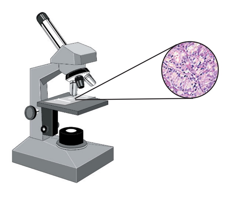
Understanding of the pathological grading is of great importance for both the clinician and the patient, as it will determine which treatment options are available, as well as their likely success. Many regional cancer centres will arrange for a specialist Pathologist to review the microscopic slides of a patient’s prostate cancer cells to confirm the diagnosis before treatment begins.